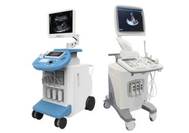
Ultrasound Systems
Individual solutions
according to your needs
Regardless of the manufacturer
What kind of ultrasound machines are you looking for?
New
Refurbished
Used
Your repair request
Get in touch with us:

Buy an ultrasound machine | A wide selection of ultrasound systems
Ultrasound is used as a diagnostic or screening tool to confirm medical impairments and to aid in performing medical procedures and treatments. By buying a used ultrasound system from a reputable trader who is specialized in sonography, considerable savings can be achieved compared to buying a new device.
Our used devices are completely overhauled and complete functional tested in our ISO 13485 certified laboratory. We offer a wide range of standard and portable systems from all manufacturers. You can choose between 2D and 3D / 4D ultrasound devices & probes for your specialty, e.g., General Ultrasound Imaging, Cardiovascular, Urology, OB / Gyn, Veterinary, Orthopedics, Pediatrics and many more.
FAQ’s Ultrasound Machines
What is ultrasound?
Ultrasound is a type of sound wave. High frequency sound waves generated by piezoelectric effects are sent back to the ultrasound machine and appear as images, called sonograms. Frequencies range from 20kHz upwards. Images are generated by the sending of pulses from the ultrasound probe (also known as “transducer”) which is placed directly inside the body (endocavitary probes – which are inserted in the vagina, oesophagus or rectum, or intraoperative probes which are used during operation in theatre). Aside from endocavitary and intraoperative probes, some probes such as linear probes, are placed directly onto the patient’s skin. The process is as follows: sound waves travel through the body, with a certain percentage being reflected to the probe and others travelling to other tissue parts. The image (sonogram) then shows the ultrasound waves which are reflected to the probe. This process is undertaken by the CPU, by which the distance from the tissue/specific organ is calculated by comparing the speed of sound (in tissue – 1540 m/s) and the time by which each echo returns. The resulting image (in 2D) is based on distances and distances and the signal intensity of the echoes which have been reflected. This is an introductory insight to ultrasonography, but there are of course more detailed explanations, which are explained below.
What are the different types of ultrasound imaging?
2-D ultrasound
The most used ultrasound imaging consists of a series of flat, two-dimensional cross-sectional images of the tissue being scanned. This type of scanning is simply known as 2-D ultrasound and, after around half a century, is still the standard for many diagnostic and obstetric situations.
3-D ultrasound
In this type of ultrasound, 2-D images are projected into three-dimensional representations. This is achieved by scanning tissue cross-sections at many different angles and reconstructing the received data into a three-dimensional image. One of the common uses for 3-D ultrasound imaging is to provide a completer and more realistic picture of a developing fetus.
4-D ultrasound
Thanks to the rapid processing of 3-D ultrasound images, sonography can also create 4-D ultrasound images. In 4-D ultrasound, the fourth dimension, time, adds movement and creates the most realistic representation of any system. In some cases, 3-D and 4-D ultrasound images can reveal anomalies that are not readily apparent with 2-D ultrasound.
Doppler ultrasound
Assessing blood flow while moving through blood vessels is a common part of many types of ultrasound. While conventional 2-D ultrasound and its three-dimensional offshoot show internal tissues and structures, another type of ultrasound is required to assess blood flow and pressure in a blood vessel. Doppler ultrasound analysis reflects high-frequency sound waves from moving blood cells and records changes in frequency in the sound waves as they return to the transducer probe. This data is then converted into a visual representation that shows how fast and in which direction blood is flowing. Doppler ultrasound is an indispensable diagnostic tool in all areas of ultrasound diagnostics and in many cases, it is preferable to X-ray angiography, as the patient does not have to be injected with a contrast media.
What are the different types of ultrasound probes?
Standard Probes
Linear – for MSK, vascular, thyroid, testes, penile, mammography, small parts, pediatrics
Convex – for obstetrics, gynaecology, abdominal, pediatrics, neonatal, kidneys, urology
Phased-Array (aka “sector-array”) – for cardiology, intracranial, some small parts, lungs
3D/4D Probes
Endocavitary 4D – for urology, gynaecology, obstetrics
Convex 4D – for abdominal, gynaecology, obstetrics, urology
Laparoscopic Probes
Intraoperative probes
TEE Probes
Transoesophageal probes – for cardiology
Are you looking for a specific ultrasound probe?
We at Ortner Medical Solutions are a trusted partner for thousands of hospitals across Europe, delivering quality, ISO 13485: 2016 certified ultrasound probe repairs, as well as probe sales. We stock probes & machines from all manufacturers and offer you excellent warranty periods and exchange discounts on your damaged ultrasound probes. Contact us today to secure your ultrasound probe deal or talk to us about our cost-effective solutions for a complete, bespoke ultrasound system.

